
Gram Staining Procedure: Step-by-Step Techniques and Accurate Interpretation
Gram Staining is a widely used and significant differential staining technique in microbiology. It was developed by Hans Christian Gram, a Danish Bacteriologist, in 1884. This test is used to distinguish between Gram Positive and Gram Negative Bacteria, aiding in the classification and differentiation of microorganisms.
Principle of Gram Staining
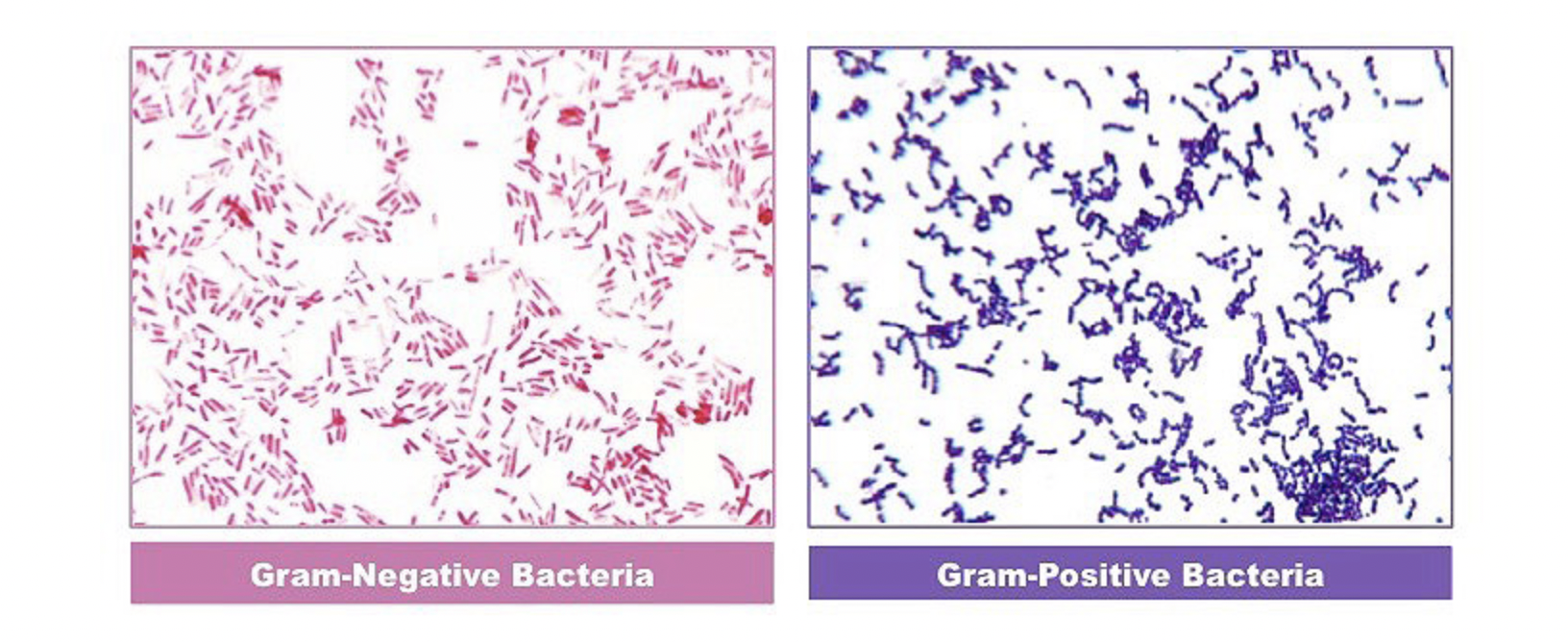
When Crystal Violet is used as the primary stain and the bacteria is fixed with a mordant, some bacteria retain the stain while others are decolorized by alcohol. Gram positive bacteria have a thick layer of peptidoglycan, which dehydrates and shrinks when the cell is decolorised. This closure of pores prevents the stain from leaving the cell, resulting in the Crystal Violet-Iodine complex remaining bound to the peptidoglycan layer and appearing blue or purple.
Gram-negative bacteria have a thin layer of peptidoglycan and a thick outer layer made of lipids. This structure causes the CV-Iodine complex to be washed off the cell wall. When exposed to alcohol, the decolorizer dissolves the lipids, allowing the crystal violet-iodine complex to leach out of the cells. After staining with safranin, the bacteria take on a red colour.
Reagents Used in Gram Staining
Crystal Violet is the primary stain, Iodine is the mordant, a decoloriser made of acetone and alcohol (95%) is used, and Safranin is the counterstain.
Procedure of Gram Staining
1.Take a slide that is clean and free of grease.
2.Use a loopful of sample to prepare a smear of suspension on the clean slide.
3.Allow it to air dry and then heat fix it.
4.Pour Crystal Violet and let it sit for approximately 30 seconds to 1 minute, then rinse with water.
5.Apply gram's iodine for 1 minute and wash with water.
6.Next, wash with 95% alcohol or acetone for about 10-20 seconds and rinse with water.
7.Add safranin for approximately 1 minute and wash with water.
8.Allow it to air dry, blot dry, and observe under a microscope.